-
CMS
Camera system
-
ORTHO
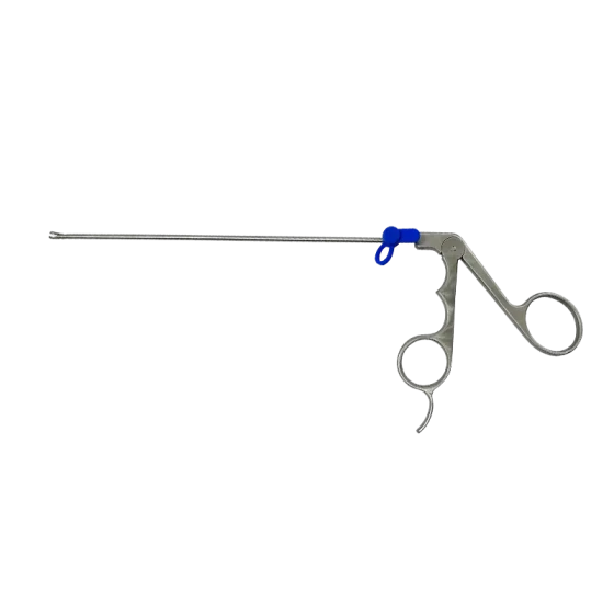
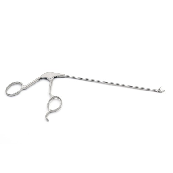
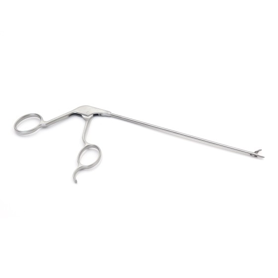
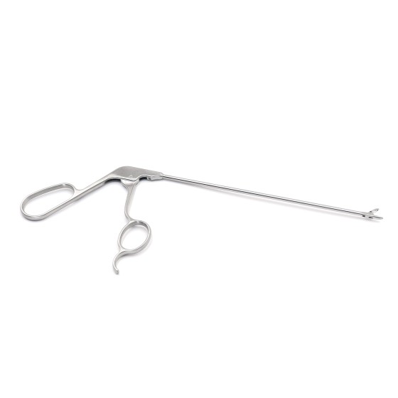
Sports MedicineSingle portal endoscopy systemDisposable Bipolar Plasma RF Electrode for OrthopedicsOrthopedic implantsOrthopedic external fixation and traction instruments
-
MUD
ResectoscopePlasma systemResectoscope operating partsElectrosurgical excision loop
- LAP
- ENT
-
MCA
Sapphire gold-platedInfusion pumpDisinfection boxElectronic endoscopeOperating room equipment
-
MB
Passive organismsPassive polymerPassive absorbable (material)Passive metal
-
News
Company NewsSpinal Surgery ColumnSpine Training Registration
- Endoscopic Spine surgery video Video
-
External fixation/implant
Implant
- Latest Products
Menu
Camera system
Sports Medicine
Single portal endoscopy system
Disposable Bipolar Plasma RF Electrode for Orthopedics
Orthopedic implants
Orthopedic external fixation and traction instruments
Resectoscope
Plasma system
Resectoscope operating parts
Electrosurgical excision loop
Sapphire gold-plated
Infusion pump
Disinfection box
Electronic endoscope
Operating room equipment
Passive organisms
Passive polymer
Passive absorbable (material)
Passive metal
Company News
Spinal Surgery Column
Spine Training Registration