Structural features and advantages
Advantages of multi-angle design
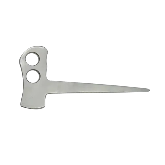
5-degree and 15-degree combination: One end of this nerve separator is at a 5-degree Angle, and the other end is at a 15-degree Angle. The 5-degree Angle is suitable for situations where the nerve is closely adhered to the surrounding tissue but requires fine separation. Due to the smaller Angle, it can be closer to the nerve surface and gently inserted between the nerve and the adhered tissue for initial separation without damaging the nerve. The 15-degree Angle is used in situations where the separation range needs to be slightly expanded or the adhered tissue is relatively tough. It can provide a greater separation force. At the same time, due to the relatively moderate Angle, it can still be precisely operated in the complex anatomical structure of the spine, avoiding damage to important tissues such as nerves. 15
The combination of 15 degrees and 25 degrees: One end of this nerve separator is at a 15-degree Angle, and the other end is at a 25-degree Angle. It is more flexible when dealing with adhesions of different degrees and various surgical scenarios. The 15-degree Angle end can be used for general nerve stripping operations, such as separating the adhesion between nerve roots and protruding intervertebral disc tissue or ligamentum flavum in spinal surgery. The end of the 25-degree Angle is more suitable for handling more complex adhesions, such as adhesions of scar tissue around nerves caused by long-term lesions. A larger 25-degree Angle can better penetrate thicker adherent tissues, helping doctors separate nerves more efficiently.
The advantages of precise operation
The designs of both nerve stripping devices are conducive to precise operations during UBE surgery. The head Angle design enables doctors to flexibly choose the appropriate Angle for stripping based on the specific conditions of the tissues around the nerves. In spinal surgery, nerve tissue is very fragile. This precision operation tool can precisely separate nerves from surrounding tissues, reducing the risk of iatrogenic nerve injury. For instance, during surgery for cervical or lumbar intervertebral disc protrusion, a nerve stripping device can precisely separate the nerve roots from the protruding nucleus pulposus tissue or the surrounding adhesive tissue along the direction of the nerve roots.
The application scenarios of UBE surgery
The application of intervertebral disc protrusion surgery
In UBE surgery for lumbar intervertebral disc protrusion or cervical intervertebral disc protrusion, a nerve stripping device is used to separate the adhesions between the nerve roots and the protruding nucleus pulposus tissue. For early-stage intervertebral disc protrusion with relatively mild adhesions, the combination of 5° and 15° nerve stripping devices can play a good role. First, use a 5° Angle end along the edge of the nerve root to separate the loose adhesions between the nerve and the nucleus pulposus. Then, as needed, use a 15° Angle end to further expand the separation range, creating favorable conditions for the removal of the nucleus pulposus. For some cases of chronic intervertebral disc protrusion with severe adhesion, the combination of 15° and 25° nerve stripping devices is more applicable. The 25° Angle end can penetrate thicker adhesive tissues, effectively separating the nerve roots from the protruding nucleus pulposus and surrounding scar tissues, and relieving the compression of the nerve roots.
The application in spinal canal decompression
In UBE surgery for spinal stenosis, a nerve stripping device can be used to separate the nerve tissue within the spinal canal from the surrounding compressive tissues, such as the ligamentum flavum and osteophytes. During cervical or lumbar spinal canal decompression, when dealing with thickened ligamentum flavum at the posterior wall of the spinal canal, a 15-degree Angle nerve stripping device (both models are available) can gently pull the spinal cord or nerve root beneath the ligamentum flavum, and then separate the adhesion between the nerve and the ligamentum flavum with an appropriate Angle of the end (depending on the adhesion condition) to prevent nerve damage during the removal of the ligamentum flavum. For lateral recess stenosis caused by osteophyte formation on the lateral wall of the spinal canal, the nerve stripping device also helps to separate the nerve from the bone, providing safety for subsequent bone removal surgery.
Application in Spinal Trauma surgery
In trauma surgeries such as spinal fractures and dislocations, nerve stripping devices can be used to examine and protect nerve tissues. For instance, in surgeries involving thoracic vertebrae fractures with a risk of spinal cord injury, nerve stripping devices can carefully examine the conditions around the spinal cord before fracture reduction and fixation, and separate bone fragments or hematoma tissues that may compress the spinal cord from it. According to the specific adhesion situation, select the appropriate Angle of the end for operation. For example, use the 15-degree Angle end for initial separation first. When encountering thicker adhesions, switch to the 25-degree Angle end to ensure the safety of the spinal cord.
Operation precautions and skills
Precautions for Operation
High requirements for visual operation: UBE surgery relies on endoscopic visual operation. Before using the nerve stripping device, it is necessary to ensure that the endoscopic field of view is clear and accurately determine the relationship between the stripping device and the surrounding nerves, blood vessels and other tissues. Any blind operation may lead to serious consequences such as nerve damage.
Gentle operation principle: Due to the fragility of nerve tissue, the principle of gentleness must be followed during operation. When inserting and using the nerve stripping device, excessive force should be avoided to prevent nerve damage. Especially when the nerve is already compressed or damaged, the operation requires even more caution.
Angle selection and conversion: The Angle of the nerve stripping head should be reasonably selected based on the degree of adhesion between the nerve and the surrounding tissues as well as the specific surgical conditions. If during the operation it is found that the current Angle is difficult to effectively separate the adhered tissue or may damage the nerve, it is necessary to carefully switch to another Angle for operation and pay attention to observing the endoscopic images to ensure the safety of the operation.
Operation skills
Insertion technique: Based on the anatomical path and nerve position observed by the endoscope, slowly insert the nerve stripping device at an appropriate Angle and direction. During the insertion process, its delicate structure should be utilized to carefully enter the area around the nerve along the natural anatomical gap or the established working channel, avoiding forced insertion that may damage the surrounding tissues. For instance, during anterior cervical spine surgery, when inserting a nerve stripping device, it can first be inserted along the gap at the anterior edge of the vertebral body, and then the direction can be slowly adjusted to approach the nerve root.
Stripping technique: When stripping the nerve from the surrounding tissue, the movement should be gentle and continuous. First, make the head of the stripping device fully contact the nerve surface or the edge of the adhered tissue, and then slowly apply the stripping force at the selected Angle. The direction of dissection should be determined based on the anatomical direction of the nerve and the requirements of the surgical operation. Generally, the separation is carried out along the natural extension direction of the nerve to avoid twisting or excessive bending of the nerve. At the same time, it is necessary to combine the real-time observation of the endoscope, and flexibly adjust the force and Angle of the dissection according to the response of the nerves and the changes of the surrounding tissues.